Tufted angioma (TA), previously known as angioblastoma of Nakagawa, is a rare, benign, vascular tumor that is thought to be on the same clinical spectrum as kaposiform hemangioendothelioma (KHE). The name describes the presence of "tufts" of capillaries in the dermis as seen on histopathological sections.
TA typically presents at birth (15%-25% of cases) or within the first 5 years of life (50% of cases) as a red-violaceous plaque or nodule. Rarely, TAs arise in adulthood. There is no consistent sex or racial / ethnic predilection.
A TA will generally grow slowly over several months to up to a decade before stabilizing in size, and partial or complete regression may occur. TAs are initially painless, but they can become painful in a significant proportion of patients.
Three clinical patterns have been described: TA without complications, TA complicated with chronic coagulopathy without thrombocytopenia, and TA associated with Kasabach-Merritt phenomenon (KMP). The mortality rate from KMP is high, from 10%-30%.
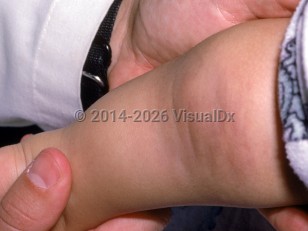
Tufted angioma
Alerts and Notices
Important News & Links
Synopsis

Codes
ICD10CM:
D18.01 – Hemangioma of skin and subcutaneous tissue
SNOMEDCT:
254786000 – Tufted angioma of skin
D18.01 – Hemangioma of skin and subcutaneous tissue
SNOMEDCT:
254786000 – Tufted angioma of skin
Look For
Subscription Required
Diagnostic Pearls
Subscription Required
Differential Diagnosis & Pitfalls

To perform a comparison, select diagnoses from the classic differential
Subscription Required
Best Tests
Subscription Required
Management Pearls
Subscription Required
Therapy
Subscription Required
References
Subscription Required
Last Reviewed:03/25/2026
Last Updated:03/25/2026
Last Updated:03/25/2026
Tufted angioma